SEM
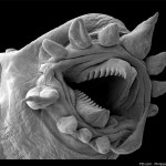
While this image may look like a creature from a science fiction movie, it's actually a hydrothermal worm marine organism viewed on an electron microscope from festival sponsor FEI Company, a world leader in the production and distribution of electron microscopes, including scanning electron microscopes (SEM), transmission electron microscopes (TEM), DualBeamâ¢Â instruments, and focused ion beam tools (FIB), for nanoscale research. See this recent Huffington Post article for more information on the image.
What microscopic organisms would you like to photograph through an electron microscope…
tags: evolutionary biology, fossils, feathers, plumage color, color, dinosaurs, theropods, Sinosauropteryx, Sinornithosaurus, birds, Confuciusornis, melanosomes, phaeomelanosomes, eumelanosomes, keratinocytes, SEM, scanning electron microscopy, 10.1038/nature08740, researchblogging.org, peer-reviewed research, peer-reviewed paper
Reconstruction of two Sinosauropteryx, sporting their orange and white striped tails.
Artwork by Chuang Zhao and Lida Xing [larger view]
DOI: 10.1038/nature08740
While looking at museum dioramas that feature dinosaurs, I often overhear people asking "How do they…
Click to Gigapan this Linepithema ant head
Gigapan is a technology that stitches together hundreds of individual images to form a massive single image. It's hard to appreciate its power from just the small SEM image shown above, but if you click on the photo you'll be able to zoom to a marvelous level of detail.
More clickable gigapan ants below the fold.
Heterospilus sp., head & compound eye, Costa Rica
Here are some shots from my training session this morning at the Beckman Institute's Scanning Electron Microscope (SEM). I haven't used SEM for years- wow! Great fun. Click on each image to enlarge.
Heterospilus sp. mesosoma
Heterospilus sp., ovipositor
For contrast, here's a photo of a wasp in the same genus taken with my standard Canon macro gear:
Heterospilus sp. Costa Rica, taken with a Canon 20D dSLR & macro lens
We'll be deciding over the coming months which type of images to use for our project.Â…
Areolate
In 1979, Rick Harris wrote a definitive paper illustrating the various terms used by taxonomists to describe the intricate patterns on the insect exoskeleton. His guide is tremendously helpful to those of us who struggle to decide if those ridges on the head of an ant are strigate or costate. Via Sifolinia, I now see that Harris's illustrations are available online:
A Glossary of Surface Sculpturing
Incidentally, Rick was the guy who taught me how to use a Scanning Electron Microscope, although at this point it'd be a minor miracle if I remembered any of it.