Did HIV become resistant to Atazanavir because of a genetic change?
Was that genetic change inherited?
Did HIV evolve?
Can we explain why genetic changes at specific sites might help HIV escape the effects of the drug?
Let's find out.
All of the sequences in the image below (except for the first) come from HIV strains that were isolated from patients who took Atazanavir and no other protease inhibitors. All of the strains of HIV from patients were resistant to the drug.
If an amino acid is different from other strains, the color at that position is changed. Since we see different shades of colors, and, if we click the image to see a larger picture, different amino acids (shown by different letters), we can conclude that the strains are genetically diverse.

Click the image to see the full-size version.
You can also see that all of the drug-resistant strains of the virus differ from the sensitive strain at two positions. At position 50, the amino acid leucine replaced isoleucine and at position 84, isoleucine replaced valine. These data show that two amino acid changes occured and were inherited by all the drug resistant isolates.
HIV evolved.
Why did the sequences change in those positions?
An interesting question, now, is what do those changes have to do with the ability of the virus to resist the drug?
Why these mutant viruses resistant to the protease inhibitor? How do they escape inhibition by the drug?
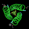
To answer this question, I downloaded a structure for a wild-type HIV protease from the Molecular Modeling Database at the NCBI. (I put a link to the structure on the Geospiza Education site, teaching materials page.) I also added annotations to the amino acids at positions, 50 and 84, to show the side chains and the amount of space that they occupy.
This structure contains a protease inhibitor (not Atazanvir) that probably binds in the same region as Atazanavir, although, it might contact different amino acids. Still, there will be some similar features. I colored this protease inhibitor pink and changed the drawing style to space fill, as well. This allows me to see how the two amino acid side chains (grey) are positioned relative to the protease inhibitor.

From the picture, it looks the two amino acids that change are located right in the binding pocket for the protease inhibitor. I think it's likely that the amino acid changes that occur at these positions prevent the drug from binding as tightly, if at all, to the viral protease. If the protease inhibitor can't bind, or binds less tightly, then it will not be able to inhibit the viral protease and the virus will be able to replicate, and infect new cells.
We've been experimenting with HIV in this series. If you want to see where we've been, here are the links.
Part I. Meet HIV and learn how we're going to use it look at evolution. An introduction to the experiment and a link to a short flash movie on HIV.
Part II. Give instructions for the experiment.
Part III. Look at the sequence results.
Part IV. Look at protein structures and see if we can explain why the experiment worked the way it did.
Reference:
1. Colonno,R., Rose,R., McLaren,C., Thiry,A., Parkin,N., Friborg,J. 2004. Identification of I50L as the Signature Atazanavir (ATV) Resistance Mutation in Treatment-Naive HIV-1Infected Patients Receiving ATV-Containing Regimens. J. Infect. Dis. 189 (10), 1802-1810.
technorati tags: digital biology,
AIDs, bioinformatics, evolution, HIV,
protease,
molecular modeling,molecular structures,
genetics