tags: evolution, evolutionary biology, UV light, flight, dinosaur, dromaeosaur, theropods, Microraptor gui, paleontology, fossils, birds, researchblogging.org,peer-reviewed research, peer-reviewed paper, journal club
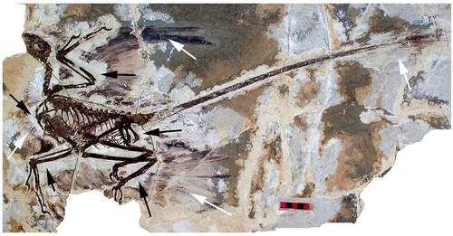
Figure 1. The holotype of Microraptor gui, IVPP V 13352 under normal light. This shows the preserved feathers (white arrow) and the 'halo' around the specimen where they appear to be absent (black arrows). Scale bar at 5 cm. [larger view]
DOI: 10.1371/journal.pone.0009223
It has long been known that when exposed to ultraviolent light, fossilized bones and shells -- and even tissues -- will fluoresce, thus rendering undetectable details visible. But this technique has been used mostly to visualize fossilized invertebrates, and inexplicably, has rarely been used to investigate hidden structures in most vertebrate fossils. But a team of paleontologists recently studied the Microraptor gui holotype using UV light.
"UV light work has been going on for decades but few people do much of it, and with the cost of color photos often only a few minor images are published, or the results are simply used to help improve a description," remarked paleontologist David Hone, a postdoctoral fellow at the Chinese Academy of Sciences, Beijing, China and first author of the newly published paper. "As such even within scientific circles this work is not too well known, something I hope will change."
Under visible light, the holotype of the non-avian dinosaur, Microraptor gui, unearthed in China, is associated with many fossilized feathers distributed around its body. In addition to numerous small contour feathers, Microraptor gui fossils also are associated with long asymmetrically shaped "flight" feathers on the arms and tail but also, surprisingly, on the legs, leading some to refer to this species as the "four-winged bird." But these flight feathers don't actually reach the limb bones in any of the dozen or so specimens of Microraptor gui examined. Instead there is a curious 'halo' of space between where the bones end and the feathers start (Figure 1, above).
''[T]here's insufficient evidence of the attachment of these feathers to the hind limbs," said paleontologist Kevin Padian at the University of California-Berkeley [DOI: 10.1641/0006-3568(2003)053[0451:FDBPON]2.0.CO;2].
"There seems to be a gap between the vaned area of feathers that are near the hind limbs and the bones of the hind limbs themselves," Dr Padian explained.
In contrast, flight feathers do reach the tail and limb bones in the basal avialan, Archaeopteryx, and in other feathered non-avian Liaoning fossils, so why is this not seen in any specimens of Microraptor gui? The obvious answer is that the fossil feathers moved from their original locations prior to preservation and thus do not appear as they did in life. They could have come free and drifted away from the bones, for instance, or the portions of the feathers that were close to the body might not have not been preserved, or they might have been destroyed during fossil preparation itself.
But regardless of the underlying reason for Microraptor gui's peculiar 'halo' effect, these specimens have meaningful implications for our understanding of the origin of flight in birds, particularly for the possibility that there might have been a four-winged gliding phase, so it is important to determine whether the fossil flight feathers were actually attached to the animal's body.
Dr Hone, together with his colleagues, Helmut Tischlinger, a retired teacher who was awarded an honorary PhD for his work with Solnhofen fossils, Xing Xu, a research fellow at the Institute of Vertebrate Paleontology and Paleoanthropology of the Chinese Academy of Sciences (CAS) in Beijing, and Fucheng Zhang, a senior research fellow at CAS, wanted to determine if the 'halo' of non-preservation observed between the skeletons and the feathery plumage is real or a preservation artifact.
To carry out this study, they spent several weeks photographing the Microraptor gui holotype while illuminating it with powerful UV-A lamps that produced light with a wavelength between 365 and 366 nanometers. They also used a variety of color correction filters (yellows, blues and reds of different types and densities and in different combinations) made from special colored glass or polyester that were affixed either to the camera or to the microscope lens (Figure 2):
Figure 2. The holotype of Microraptor gui, IVPP V 13352 under UV light. Different filters were employed for parts A [larger view] and B [larger view], hence the difference in colour and appearance. A also is labeled to indicate the preserved feathers (grey arrows) and the 'halo' around the specimen where they appear to be absent (black arrows) as well as phosphatised tissues (white arrows). Scale bars are 5 cm in both A and B.
DOI: 10.1371/journal.pone.0009223
"The mechanics are fairly simple in that we basically just shine some very powerful UV lights at the fossil and take photos," explained Dr Hone. "It can take dozens of shots to get a good one and some of the exposures take an hour or more. It therefore takes a lot of time, skill, patience and especially experience to get these results."
Using this method, the team focused on the hind limbs and found that shafts of the UV-illuminated feathers are visible in the 'halo' region (Figure 3):
Figure 3. Close up of lower hindlimb of the holotype under UV light. This shows that the feathers do indeed penetrate the halo (grey arrows) when seen in UV and approach or reach the bones. These are not seen in natural light due to the overlying soft tissues seen in figure 2. Scale bar at 5 cm. [larger view]
DOI: 10.1371/journal.pone.0009223
"[T]he fact that the leg feathers do have bone-deep attachments could be used as an argument that they are used in flight," continued Dr Hone. "However, the asymmetry of these feathers is a far more important character and we can't really add anything to the previous work in that respect."
Further, the feather filaments around the throat and chest were also quite clear under UV illumination when in fact, they are invisible under natural light (Figure 4):
Figure 4. Close up of the chest of the holotype, close to the sternal plates under UV light. As with figure 3, this shows that the feathers do indeed penetrate the halo (grey arrows) when seen in UV and approach or reach the bones. These are not seen in natural light due to the overlying phosphatised tissues, but the striations of the feathers are clearly visible despite this covering. Scale bar of 1 cm. [larger view]
DOI: 10.1371/journal.pone.0009223
Based on their findings, Dr Hone and his team concluded that Microraptor gui's feathers are probably preserved in their original and natural positions. The feather were not decayed, nor were they moved or otherwise disturbed prior to fossilization. Second, they also found that Microraptor gui's feathers do actually reach the bones of the animal, even though their bases were obscured. Of course, it also means that previously published measurements of the feathers are in error: the feathers are longer than assumed.
Figure 5. Close up of the lower part of the holotype under UV light. Variation in the phosphatised tissues can be seen (white arrows) as well as the bright reflectance of various glues and preservatives that have been applied to the specimen at various times (black arrows). Scale bar of 2 cm. [larger view]
DOI: 10.1371/journal.pone.0009223
Even though the findings reported in this paper provide a nice opportunity to show you lots of detailed photographs of Microraptor gui, the take-home message is about what UV illumination means in regards to fossil preservation and preparation. There is a lot of hidden information waiting to be uncovered in many fossils, even long after they've come to light, and this information can be accessed by using a variety of techniques, such as UV light illumination.
Unfortunately, scientists miss all kinds of rare and valuable soft-tissue information by not using all of the techniques available to them, often because they are unaware of them. If paleontologists and fossil preparators are unaware of the variety of hidden information that a fossil might contain, they risk destroying it during preparation. Already, at least one museum now regularly prepares fossil material with UV light (as already has happened with one famous fossil, Juravenator, for example).
"This lack of awareness is certainly starting to change with UV work slowly gaining attention and prominence but I hope that this paper will accelerate that process," Dr Hone writes on his blog, Archosaur Musings.
Dr Hone and his team hope that publishing their study in PLoS ONE will raise paleontologists' awareness of the utility of this powerful technique.
"PLoS1 is, after all, an open access journal and UV stuff is not especially hard to do. Now that there is extensive documentation of the methods in a very well distributed format, that can only be a good thing bearing in mind the results that UV can bring."
Dr Hone and his colleagues also think that taking another, closer look at well-studied fossils using UV illumination could yield more discoveries that have remained hidden under the light of day.
"Long may [UV illumination] continue, and more importantly, the faster it spreads the more chance we have of being able to write papers like this in the future where we can get new information out of otherwise 'exhausted' specimens."
Source:
Hone, D., Tischlinger, H., Xu, X., & Zhang, F. (2010). The Extent of the Preserved Feathers on the Four-Winged Dinosaur Microraptor gui under Ultraviolet Light. PLoS ONE, 5 (2) DOI: 10.1371/journal.pone.0009223


There's an interesting series on the Beeb at the moment about the Natural History Museum in London, and all the stuff they do besides the public galleries. Part of that was about the massive collection of specimens that people don't normally see, kept for reference and for further study with new techniques.
I find it fascinating that people can learn so much from such complicated and obscure things as fossils and microscopy, just by looking at them. I had enough trouble doing cell drawings at school when I knew what I was looking at. (Total absence of artistic talent didn't help.)