Over 2600 genetic diseases have been found where a change in a single gene is linked to the disease. One of the questions we might ask is how those mutations change the shape and possibly the function of a protein?
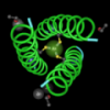
If the structures of the mutant and wild type (normal) proteins have been solved, NCBI has a program called VAST that can be used to align those structures. I have an example here where you can see how a single amino acid change makes influenza resistant to Tamiflu®.
This 4 minute movie below shows how we can obtain those aligned structures from VAST and view them with Cn3D.
CN3D and VAST from Sandra Porter on Vimeo.
I love this! If only more mutant structures had been solved, this could really help my work.
Thanks Rob!
You'd be surprised, I think, at how many structures are available! Every day, I find new ones in the database.